
As such, these well-defined gels provide promising versatile matrices for fostering elaborate in vitro morphogenesis. These tunable matrices are stress relaxing and thus promote efficient crypt budding in intestinal stem-cell epithelia through increased symmetry breaking and Paneth cell formation dependent on yes-associated protein 1. Here we report a family of synthetic hydrogels that promote extensive organoid morphogenesis through dynamic rearrangements mediated by reversible hydrogen bonding. Chemical or enzymatic degradation schemes can partially alleviate this problem, but due to their irreversibility, long-term applicability is limited. However, the excessive forces caused by tissue expansion in such elastic gels severely restrict organoid growth and morphogenesis. Efforts to replace such ill-defined matrices for organoid culture have largely focused on non-adaptable hydrogels composed of covalently crosslinked hydrophilic macromolecules. General implications for the size measurements of polymer nanoparticles in solutions are discussed.Įpithelial organoids are most efficiently grown from mouse-tumour-derived, reconstituted extracellular matrix hydrogels, whose poorly defined composition, batch-to-batch variability and immunogenicity limit clinical applications. Basing on results of our polysilane measurements, we come to a conclusion that DLS provides more reliable results than SEC for the dilute solution of globules. The only possibility to measure aggregation-free, a true molecular size of polymer nanoparticles in such regime of solution, is to operate with the dilute solution of globules (below theta point and above the miscibility line). Determination of particle sizes in presence of attractive interactions is not a trivial task. Polymer nanoparticles are typically in poor solution conditions below the theta point and are in globular conformation therefore. Taking dendritic and network polysilanes as a group of least soluble polymer substances, we critically compare and discuss the difference between nanoparticle sizes, obtained by DLS and SEC.

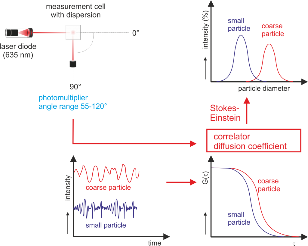
Owing to its simplicity and low-sample volume requirements, DLS can be used even by hospital pharmacists to confirm absence of protein aggregates immediately before drug administration.Dynamic Light Scattering (DLS) and Size Exclusion Chromatography (SEC) are among the most popular methods for determining polymer sizes in solution. Although no single techniques can reveal all aspects of protein stability, DLS can serve as a screening tool to detect aggregate formation and cross-validate SE-HPLC results during batch release testing. Results showed that aggregate formation was not detected in some cases by SE-HPLC and the decrease in the concentration of the monomeric forms indicated that such aggregates might have been filtered off the column. Generally, good agreement between the results of DLS and SE-HPLC was noted, regardless of the differences in physicochemical properties of the studied biopharmaceuticals. Samples were subjected to forced degradation conditions previously shown to lead to aggregate formation (pH 4.0, 8.0 and 10.0, at 37 ☌ for 24 h) and samples were analyzed using DLS and SE-HPLC. Here, three model biopharmaceutical proteins of different physicochemical properties were selected quadrivalent human papillomavirus virus like particles vaccine (HPV VLP, physically assembled subunit vaccine, 55 kDa), pegylated Interferon (PegIFN, pegylated non-glycosylated protein, 31.3 kDa) and Pegylated Erythropoietin (PegEPO, pegylated and glycosylated protein, 60 kDa). On the other hand, dynamic light scattering (DLS) is a simple and non-destructive technique that can detect high molecular weight physical and chemical aggregates in their native environment. Moreover, low-affinity protein aggregates may dissociate during analysis and thus not detected. However, large protein aggregates may be filtered off and build up on top of the column leading to deterioration in column performance. Size exclusion high performance liquid chromatography (SE-HPLC) has always been the gold standard technique for detection and determination of protein aggregates.

Aggregate formation is a major problem affecting both safety and efficacy of biopharmaceuticals and is associated with protein immunogenicity.


 0 kommentar(er)
